|
SAA: Serum amyloid A
Serum amyloid A (SAA) is a type of protein in the blood that is secreted by the liver. For a healthy individual, SAA concentration in the blood is usuallylow; however, in the case of inflammation or malignant tumor formation, the level of serum SAA would significantly increase, possibly as a part of the liver’s non-specific response to the inflammation or tumor. However, reports of SAA overexpression in cancer tissues aside from the liver indicate that cancer cells can also secrete SAA. This is assumed to be driven by the immunosuppressive effect of SAA, which gives the SAA-expressing tumor cells competitive advantage in the body.Since 1984, SAA has been identified to be closely related to malignant tumors. In many studies, SAA was observed to be involved in the formation and progression of several types of cancers, including lung cancer, colorectal cancer, prostate cancer, nasopharyngeal carcinoma, pancreas cancer, and breast cancer. In some studies, the concentration of SAA in the blood wasused as a diagnostic tool. For patients with colorectal cancer metastasis, the increase of serum SAA concentration is closely related to the disease. In addition, a new variant (SAA-HN) was found in hepatocellular carcinoma, particularlyin those with late stage alcohol-related liver disease such asliver cirrhosis. The clinicopathological characteristics of SAA-HN are highly related to itspotential to become a liver cancer cell.
In what ways can SAA be used to detect cancers?
Although there are a lot of evidence demonstrating the close relationship of SAA and cancer, SAA cancer detection toolsare not yet available on the market. This is because previously, the researchers could only screen for the total amount ofSAA, which are not sufficient to distinguish those with high risk of cancer from those that only have inflammation.
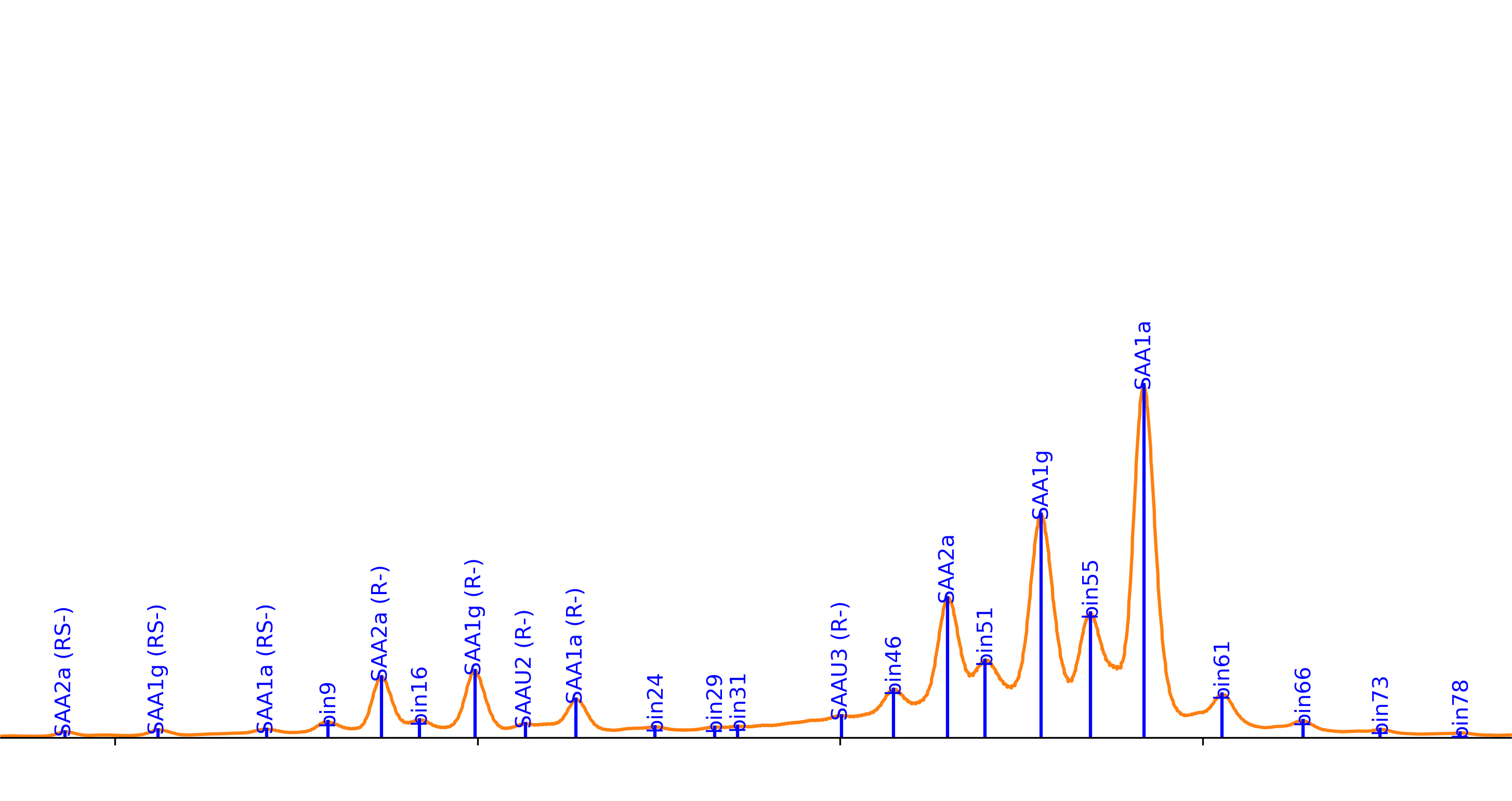
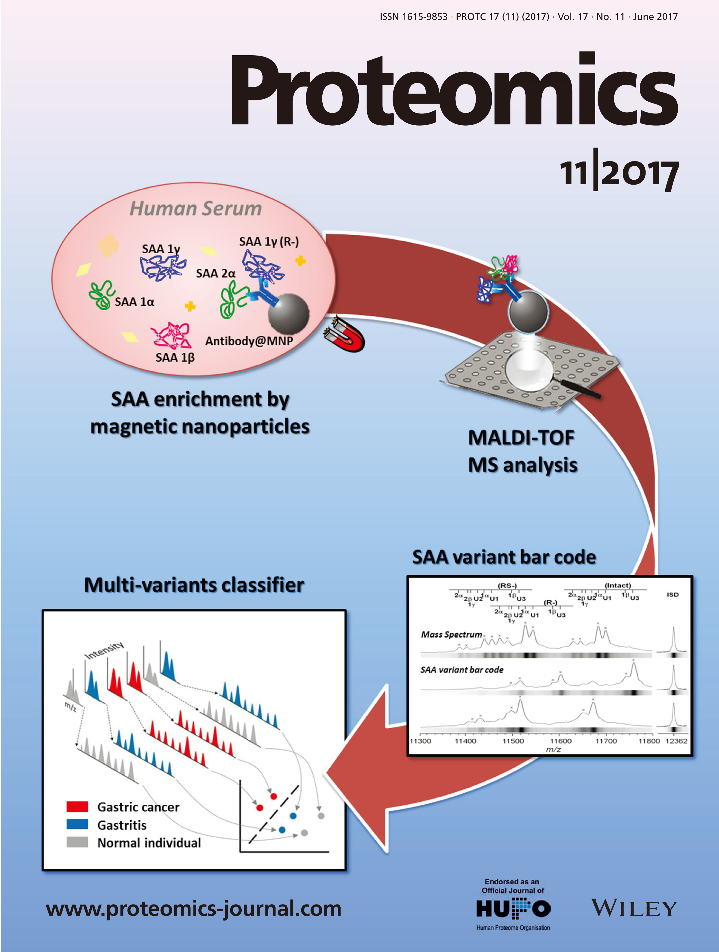
Based on previous reports, there are over 24 types of SAA isoforms. These arise from allelic variants, and N- and C- terminal truncations.Depending on the condition of the cancer patient, thecomposition and relative amountsof the different isoformsmay change. Existing immunoassays such as ELISA are unable to distinguish these different isoforms. Mass spectrometers are currently the only instrumentsable to clearly differentiate the various SAAisoform pattern and in turn, correlate them todisease status.
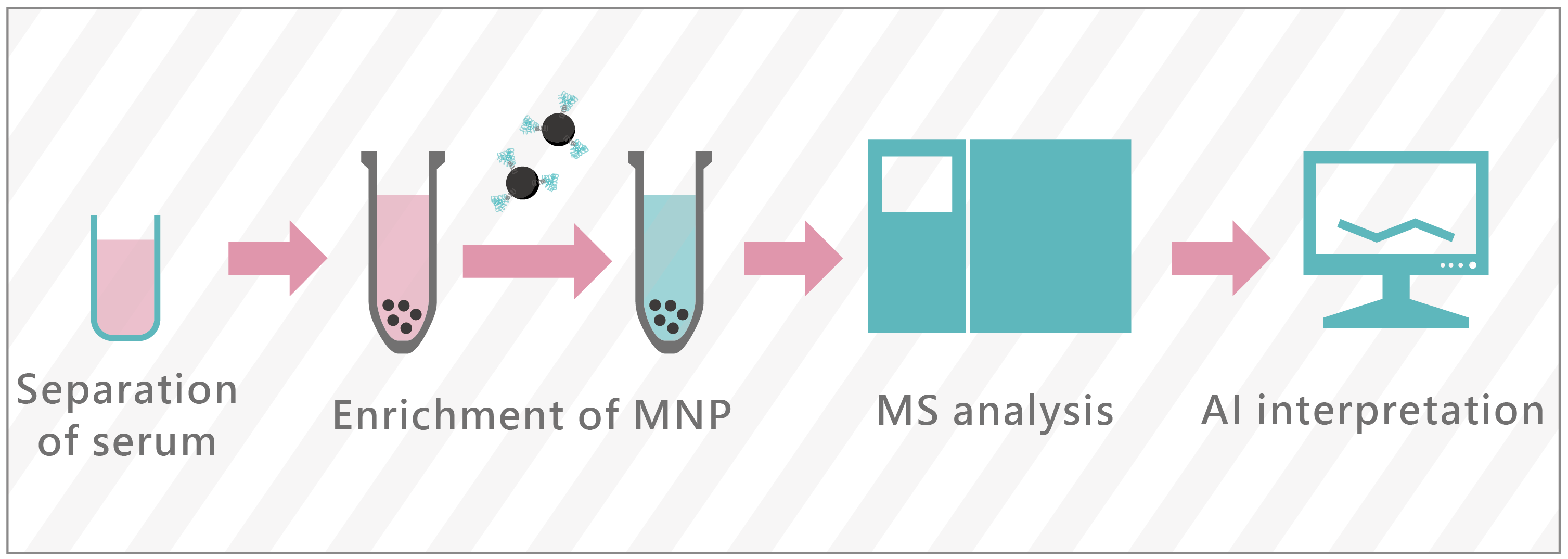
Adding to the challenge is the low concentration of SAA in the blood, which makes it difficult to bepurified and examined. Our cancer detection test utilizes magnetic nanoparticles to purifySAA, employs a MALDI-TOF mass spectrometer for rapid detection of the isoforms, and finally uses AI technologyto identifythose with high risk of cancer.
Based on previous reports, there are over 24 types of SAA isoforms. These arise from allelic variants, and N- and C- terminal truncations.Depending on the condition of the cancer patient, thecomposition and relative amountsof the different isoformsmay change. Existing immunoassays such as ELISA are unable to distinguish these different isoforms. Mass spectrometers are currently the only instrumentsable to clearly differentiate the various SAAisoform pattern and in turn, correlate them todisease status.
Adding to the challenge is the low concentration of SAA in the blood, which makes it difficult to bepurified and examined. Our cancer detection test utilizes magnetic nanoparticles to purifySAA, employs a MALDI-TOF mass spectrometer for rapid detection of the isoforms, and finally uses AI technologyto identifythose with high risk of cancer.
|
The technology authorized by many top research and development teams in Taiwan.
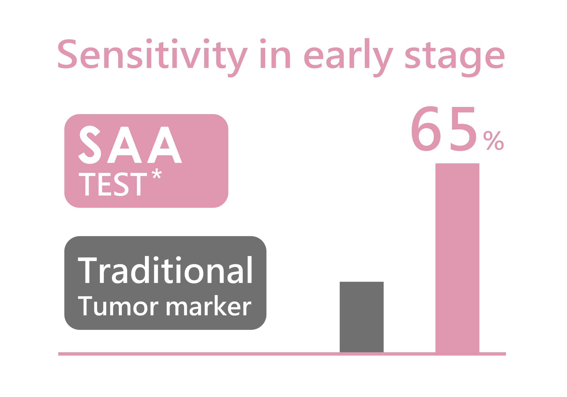
Technologies obtained technology transfer from multiple academic and research institutions with years of research and development, the SAA TEST has been done. The technology of SAA MS screening and interpretation of MS spectra come from academic and research institutions. NTHU is responsible for the development of Magnetic Nanoparticles and antibody for extraction and purification of SAA. NYMU and Metal Industries Research & Development Centre help develop the bio-algorithms, making it more accurate. Last but not least, KMU work on clinical trials and the integration of clinical information to ensurethat SAA nano MS screening service has high accuracy and also help practically in the medical field.The strongest advantage: 65%
of sensitivity in early stage of cancer
The biggest difference between SAA TEST and other cancer screening service is the detect ability factor. SAA TEST has 65% of sensitivity in early stage of cancer.
Accumulated and advanced and research findings
|
|